
Health
Cancer Immunotherapy: The Role of Tertiary Lymphoid Structures (TLS)

On this page
By Pierre-Alexis Da Costa, PhD candidate in the Inflammation Complement and Cancer team at the Centre de Recherche des Cordeliers, supervised by Prof. Isabelle Cremer
The identification of prognostic biomarkers in cancer represents a key challenge for improving patient monitoring and care. The ability of the immune system to fight cancer cells is one of the most critical factors in the anti-tumour response. Among immune cells, lymphocytes emerge as a key prognostic marker, particularly when they organize into aggregates called tertiary lymphoid structures (TLS).
However, the presence and number of TLS vary considerably from one patient to another. The mechanisms underlying TLS formation remain insufficiently understood: it is therefore essential to better characterize them to optimize immunotherapy treatments.
In this article, you will learn about the nature of TLS and its clinical significance.
The Tumour and Its Microenvironment
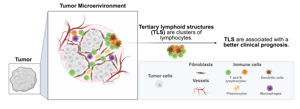
A tumour is not simply a mass of cancer cells: it evolves within a complex, heterogeneous, and dynamic ecosystem called the tumour microenvironment. This microenvironment encompasses all elements surrounding the tumour, including tumour cells, blood vessels, fibroblasts, and immune cells (Swartz et al., 2012).
Within this microenvironment, immune cells perform different roles depending on their type. Lymphocytes, also known as white blood cells, play a central role in the body’s defence against the tumour. Two main groups can be distinguished:
• T lymphocytes mediate the cellular immune response by directly destroying tumour cells or coordinating the immune response.
• B lymphocytes provide humoral immunity by producing monoclonal antibodies directed against the tumour.
Under chronic inflammatory conditions, these lymphocytes can organize into structured aggregates called tertiary lymphoid structures (Fridman et al., 2022).
Understanding Tertiary Lymphoid Structures (TLS)
Organized Lymphocyte Aggregates
TLS were named by analogy with primary and secondary lymphoid organs:
• Primary lymphoid organs (bone marrow, thymus) are sites of lymphocyte maturation.
• Secondary lymphoid organs (lymph nodes, spleen) serve as sites for immune response activation, notably through dendritic cells that enable lymphocyte activation.
TLS are distinguished by their ectopic location within the tumour microenvironment. They correspond to organized lymphoid structures that develop outside classical lymphoid organs and constitute local sites of interaction between different immune cells.
TLS can be compared to a local immune “barracks,” where lymphocytes gather, organize, and specialize to more effectively coordinate the immune response directed against the tumour (Gago Da Graça et al., 2021).
TLS Formation Involves Numerous Molecular Actors
TLS neogenesis involves various processes governed by numerous factors. Among the identified mechanisms:
• Local production of secreted molecules, notably cytokines and chemokines (lymphotoxin-α1β2, CCL19, CCL21, CXCL13), contributing to lymphocyte attraction and recruitment (Aloisi & Pujol-Borrell, 2006).
• The presence of specialized vessels, such as high endothelial venules and lymphatic vessels, facilitating lymphocyte infiltration within the tumour.
• The involvement of specific cell populations: dendritic cells, follicular dendritic cells, follicular helper T lymphocytes (Tfh), and fibroblastic reticular cells (FRC), participating in TLS organization and maintenance (Fridman et al., 2022).
Different Stages of TLS Maturation
Depending on their level of organization, TLS present different maturation stages (Fridman et al., 2023):
Immature TLS Aggregates of T and B lymphocytes lacking well-defined separate zones.
Mature TLS are classified into two categories:
• Primary Follicle-Like TLS (PFL-TLS): characterized by the presence of follicular dendritic cells and Tfh lymphocytes, enabling B lymphocyte activation and initiation of the humoral response.
• Secondary Follicle-Like TLS (SFL-TLS): characterized by the presence of a germinal centre allowing B lymphocyte differentiation into plasma cells secreting high-affinity antibodies directed against cancer cells (Fridman et al., 2022).
TLS maturation likely depends on persistent antigenic stimulation and continuous production of chemokines and cytokines (Peyraud et al., 2025).
TLS Detection in Research and Hospital Settings
TLS can be detected using various methods.
Detection Techniques
TLS can be detected on histological sections stained with haematoxylin, eosin and safranin (HES), a technique commonly used in clinical practice. Their identification is now facilitated by recently developed artificial intelligence algorithms (Vanhersecke et al., 2022; Chen et al., 2024).
In research, TLS identification also relies on immunohistochemical staining targeting:
• T lymphocytes (CD3)
• B lymphocytes (CD20 or CD19)
• Follicular dendritic cells (CD21 and CD23)
CD23 expression by follicular dendritic cells indicates the presence of a germinal centre and thus allows identification of mature TLS.
Finally, TLS can also be studied through transcriptomics (RNA sequencing), using various gene signatures to estimate their quantification (Meylan et al., 2022; Wu et al., 2021).
A standardized method for TLS detection, classification, and maturation assessment has recently been proposed, facilitating their characterization in routine clinical practice (Vanhersecke et al., 2023). Application of this algorithm revealed the presence of mature TLS in 33.5% of cases in a cohort of 1,393 patients (Brunet et al., 2023).
TLS plays a key role in the anti-tumour response
The presence of TLS is associated with a better prognosis and a better response to immunotherapy treatments in many cancers, including lung, kidney and breast cancers (Bouloudani et al., 2025).
However, this beneficial effect is mainly observed in patients with mature TLS. (Petitprez et al., 2020; Vanhersecke et al., 2021)
Clinical and Therapeutic Perspectives
The biological mechanisms responsible for the formation and maturation of TLS are still not fully understood. Current research focuses on two main areas (Peyraud et al., 2025):
• Improving TLS detection.
• Developing personalized therapeutic strategies capable of inducing mature TLS formation to strengthen the anti-tumour immune response.
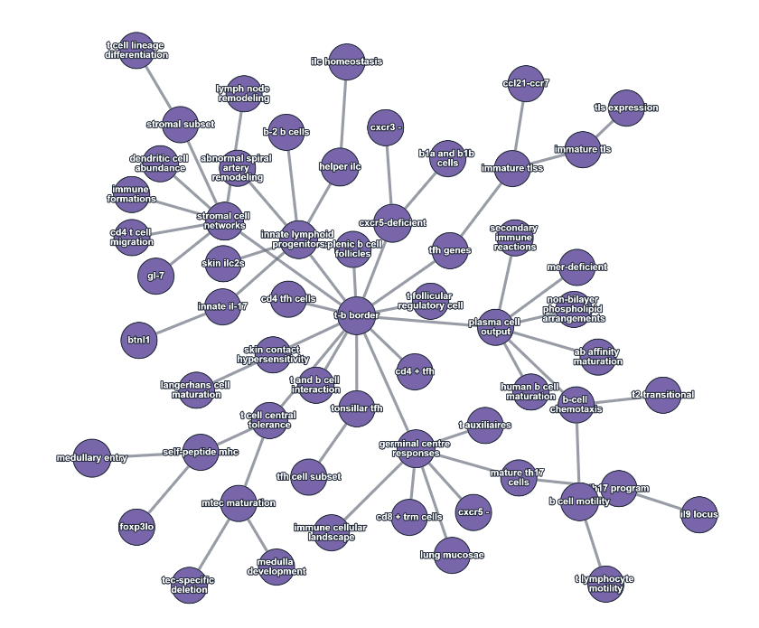
Knowledge graph on tertiary lymphoid structures, generated using NeoPhi. This graph maps the key concepts, relationships, and research findings from peer-reviewed literature on tertiary lymphoid structures and their role in immune response.
FAQ: Immunotherapy and Tertiary Lymphoid Structures
What is a tertiary lymphoid structure (TLS)?
TLSs represent a spatial organisation of lymphocytes and can be likened to a local immune ‘barracks’, within which lymphocytes congregate, organise themselves and specialise in order to better coordinate the response against the tumour.
What are cytokines?
Cytokines are proteins that play a vital role in immune regulation by coordinating inflammatory responses. Among cytokines are chemokines, which act as chemoattractants, guiding immune cells to sites of inflammation.
What is immunotherapy?
‘Immunotherapy works by stimulating a patient’s immune system to help it fight the disease. In the case of cancer, it does not attack the tumour directly, but stimulates the immune cells involved in recognising and destroying tumour cells.’ Source: https://www.inserm.fr/dossier/immunotherapie-cancers/
Bibliography
Keep reading
From Bench to Pipeline: Self-Supervised Learning for Noisy Biomedical Signals in Pharma and Biotech R&D

Dietary supplements and ADHD : What does the scientific literature really say about omega-3s, saffron, and micronutrients?